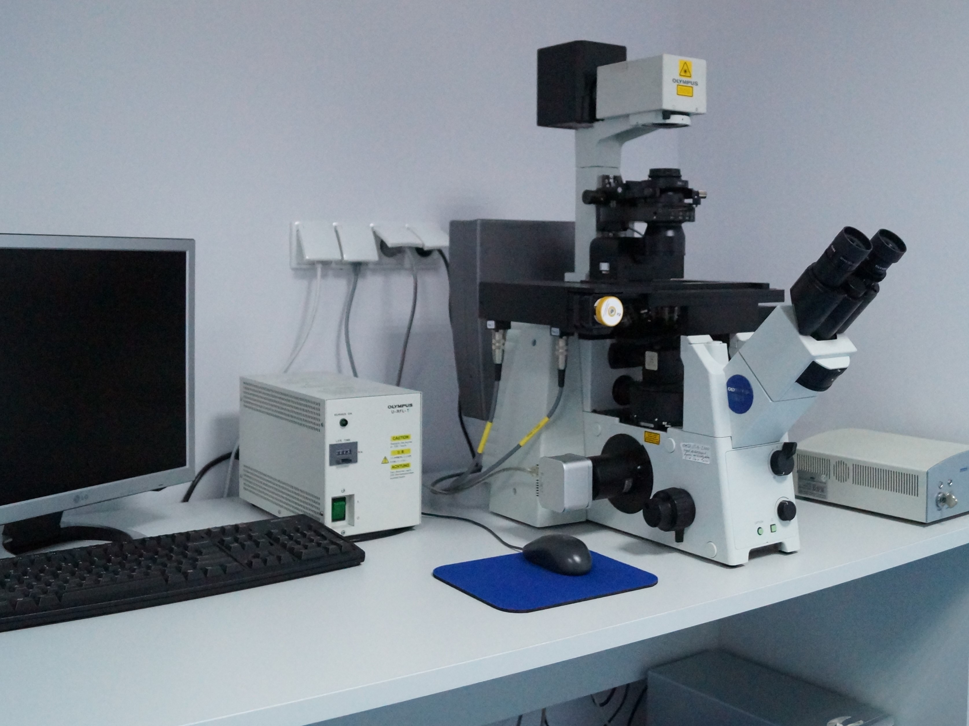
1. Laser Microdissection System Cell Cut Plus (MMI) with IX71 inverted fluorescence microscope (Olympus).
The CellCut system offers precise and reliable laser microdissection/ micromanipulation for molecular biology, cytogenetic, molecular medicine, pathology, and forensic medicine applications. This system requires using cultures dishes or slides covered with a special membrane intended for this type of equipment.
• Inverted microscope IX 71 (Olympus) equipped with objectives: U Plan-FLN x4.0,13, U Plan-FLN x10/0,3, LUC Plan-FLN x20/0,45, LUC Plan-FLN x40/0,60,
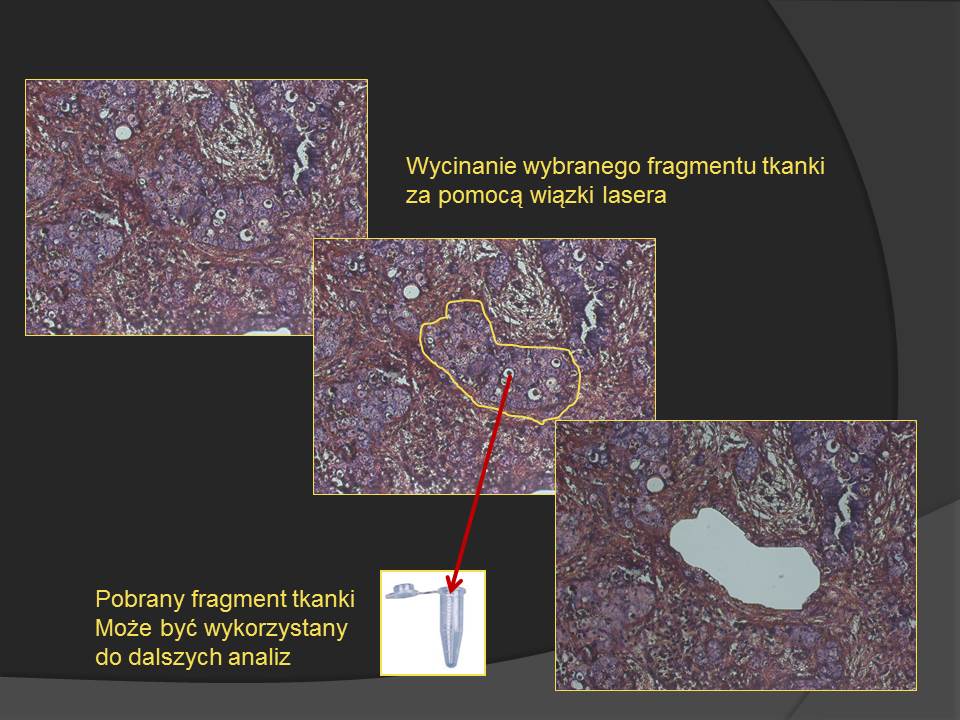
• The laser emits a light beam with a length of 448 nm, which enables precise contouring and cutting (microdissection) of selected cell fragments from cultures or tissues from histological, immunohistochemical, and immunofluorescent specimens of standard thickness,
• Selected, single, or multiple samples can be collected in sterile tubes using the system based on the patented contamination-free membrane and CapLift technology
• The progressive scan CCD delivers outstanding image quality, which – in conjunction with the intuitive UVCut Plus software – facilitates the precise drawing of cutting paths into the live image.
Software: MMI CellTools with UVCut Plus plug-in – v.3.47
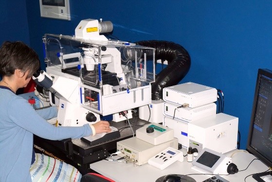
2. System for long-term in vivo observations - Cell Observer SD (Zeiss)
The system is designed to observe and register changes occurring over time, including migration, chemotaxis, or dynamic measurements of parameters such as calcium concentration, pH, and mitochondrial membrane potential changes.
• Axio Observer Z.1 inverted microscope with an incubation chamber with temperature, humidity, CO2, and O2 regulation (hypoxic conditions possible) dedicated for long-term in vivo imaging in Petri dishes (diameter 3.5-5.0 cm), Lab-Tek, and in multi-well dishes (6, 24, 96).
• Modified Spinning Disk system (Yokogawa modification) with two optical paths and two EM CCD (dual camera) cameras, and a laser module (4 diode lasers - 405 nm, 488nm, 561nm, 635nm) enable high-speed imaging in the semiconfocal mode.
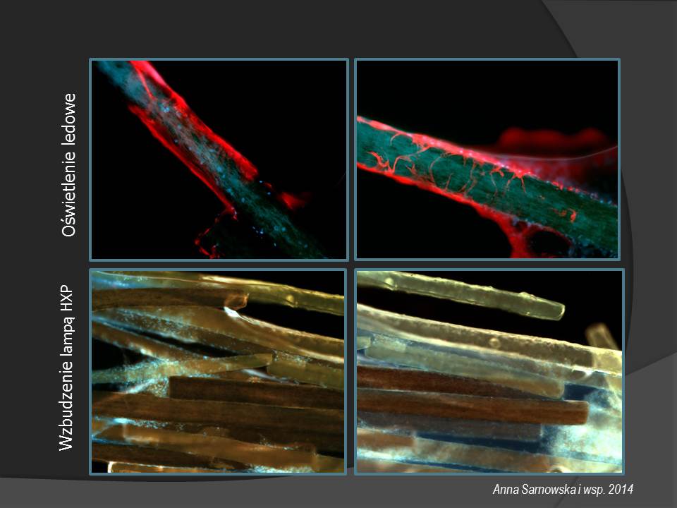
• Colibri.2 lighting system - a set of LED illuminators: 365nm, 470nm, 540-580nm, 590nm enables fast fluorescent imaging of cells, organotypic sections, and tissues as well as 3D structures (organoids, scaffolds) in the plane (XY) and along the axis ( With).
• HXP 120V fluorescent lamp, third AxioCam MRm camera for recording standard fluorescence,
• Definite Focus v.2 system,
• Objectives: standard or LD for observing specimens on slides, multi-well dishes, or a culture dish with a glass bottom. EC Plan-Neofluar 10x/0.30 M27, Plan-Apochromat 20x/0.8, LD Plan-Neofluar 20x/0.4 Korr, C-Apochromat 40x/1.2 W Korr, LD Plan-Neofluar 40x/0.6 Korr, Plan-Neofluar 63x/1.4 Oil DIC.
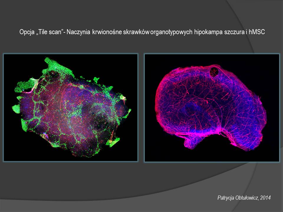
Software: ZEN 2012 blue edition and additional modules: Tiles/ Positions and Experiment Designer modules, High Content Analysis – ASSAYbilder.
3. Confocal microscope with manual stage - LSM 510 (Zeiss)
The system with a manual stage enables examinations at the level of tissues, cells, and cellular structures, such as simultaneous registration of 4-6 fluorescent signals in vivo systems and fixed specimens, registration of images in the (xy) and (z) axis, long-time observations, reconstructions 2, 5D and 3D, ortho projection, as well as physiological studies and dynamic measurements of calcium concentration, pH, changes in mitochondrial membrane potential
• Axiovert 200M inverted microscope, additionally equipped with a small incubation chamber for long-term observation, under constant conditions of temperature (heated stage), CO2 concentration, and humidity,
• 4 lasers (diode 405nm, HeNe 543nm, HeNe 633nm, Ar multi-line 458/477/488 / 514nm),
• Objectives: Plan-Neofluar 10x/0.30 Pol, Plan-Neofluar 20x/0.5, Plan-Neofluar 40x/1.3 Oil DIC, Plan-Apochromat 63x/1.40 Oil DIC M27,
• Simultaneous 3-channel fluorescence and DIC imaging
• AxioCam HRm Rev.2 digital camera.
Software: Zen 2011 black edition. Standard software for colocation measurements.
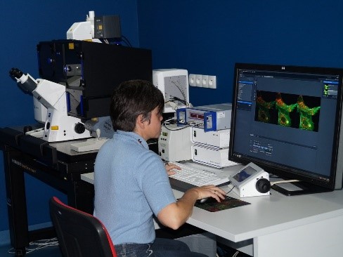
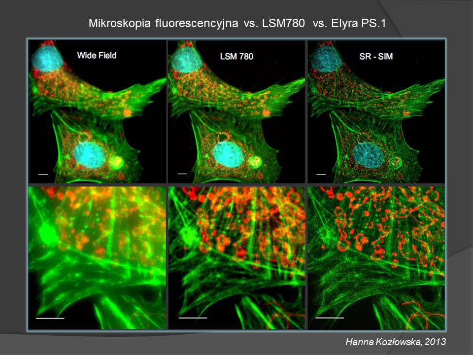
4. Confocal microscope with automatic stage, spectral, and super-resolution modules - LSM 780 / ELYRA PS.1 (Zeiss)
The system combined the confocal spectral detection LSM780 and ELYRA PS.1 head with two methods of super-resolution: PAL-M and SR-SIM.
The device can be used to study the expression of cellular antigens, cell morphology, and tissue structure and register labeled cell organelles, e.g., mitochondria and extracellular matrix.
The research concerns fixed cell cultures, tissues, or organotypic sections - multichannel imaging, signal co-localization, cell division, movement, measurements of calcium ion concentration changes, monitoring of potential membrane changes.
The use of sensitive detectors makes it possible to minimize the power of lasers used during the registration of images from fixed preparations. It enables simultaneous registration of the total signal spectrum and precise spectral separation of overlapping signals, including tissue autofluorescence. In addition, it allows you to measure weak signals. The LSM 780 confocal system is equipped with a GaAsP spectral detector. Since this is a parallel spectral detection design, it offers a simultaneous 34-channel readout in lambda mode. Up to 10 dyes can be acquired and separated at the same time. Additionally, it allows the measurement of weak signals and counts individual photons.
• Axio Observer Z.1 inverted microscope,
• Objectives: EC Plan-Neofluar 10x/0.30 M27, Plan-Apochromat 20x/0.8, Plan-Apochromat 40x/0.95 Corr M27, Plan-Apochromat 63x/1.40 Oil DIC M27, Plan-Apochromat 100x/1.46 Oil DIC M27, Plan-Apochromat 100x/1.57 Oil DIC Korr M27.
• confocal spectral detection module with 34 detectors, 4 lasers (405nm diode, 561nm solid-state laser, HeNe 633nm, multi-line Ar 458/488 / 514nm),
• ELYRA PS.1 super-resolution module with 4 lasers (405nm diode, 561nm solid-state laser, 488nm solid-state laser, 642nm solid-state laser), two EM CCD-cooled cameras for super-resolution recording.
ELYRA PS.1 combines two super-resolution recording techniques in one head: SR-SIM (Superresolution Structured Illumination Microscopy) and PAL-M (Photo-activated Localization Microscopy), which give the application versatility of the research. SR-SIM enables the registration of images at four excitation wavelengths (with the possibility of overlapping channels) with a resolution of approx. 100 nm in the (x,y) plane and 200 nm in the (z) plane for preparations stained as imaging in a traditional fluorescence microscope. PAL-M for labeling requires proteins that can be photoactivated or photoconverted. The resolution obtained with this method is approximately 20 nm in the (x, y) plane and 100 nm in the (z) plane.
Software: Zen 2012 black edition and additional modules: Tiles/ Positions and Experiment Designer, Photoactivated Localization Microscopy, Structured Illumination.